It’s easy to ignore a sore spot on your tongue or other areas inside your mouth and hope it heals on its own. But, if you can’t link it to any specific cause like a burn, trauma, or abrasion, it’s probably a good time to drive down to your dentist’s office for a check-up.
These red patches may be erythroplakia, a precancerous condition with a high probability of becoming oral cancer if left untreated. Learning about erythroplakia symptoms and causes can help reduce your chance of developing oral cancer.
What Is Erythroplakia?
Erythroplakia is a rare, painful, slow-growing, red or purple lesion on the mucous membranes of the mouth. They can be flat or raised, with distinct margins. Other erythroplakia symptoms include bleeding when scraped. Along with leukoplakia lesions — white or gray patches on the tongue or mouth’s lining — erythroplakia is usually found on the floor of the mouth or the tongue.
Leukoplakia and erythroplakia are considered potential precancerous conditions of the mouth. Leaving them untreated allows them to progress to oral cancer by undergoing malignant transformation. According to a study published in 2016, erythroplakia’s malignant transformation rates range from 14% to 50%, with a high chance of having precancerous cells.
While not all patches in the mouth are cancerous or precancerous, visit your dentist or doctor for a diagnosis to ensure that the patches in your mouth are harmless.
Erythroplakia: Causes
Heavy smoking, tobacco, and excessive alcohol consumption are the usual erythroplakiacauses. Other risk factors include poor oral hygiene, long-term trauma to the mouth due to ill-fitting dentures, and human papillomavirus infection.
How Is Erythroplakia Diagnosed?
Early detection and diagnosis are important. Some patients might not experience erythroplakia symptoms, increasing their risk of going unnoticed and becoming oral cancer. Some methods used to diagnose erythroplakia include:
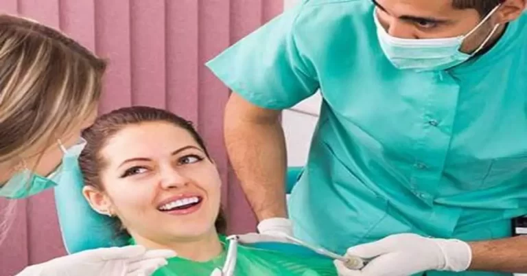
Visual Inspection
A healthcare provider, often a dentist, can diagnose erythroplakia by looking at the mouth. They may also use a thorough medical history and review a patient’s signs and symptoms to rule out other conditions.
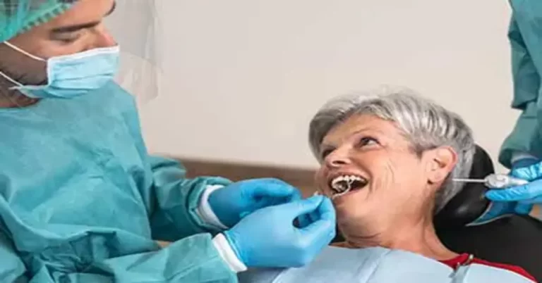
Biopsy
Your dentist or doctor may also recommend a biopsy to confirm a diagnosis. It involves taking a sample from the lesion and examining it in a laboratory. Technicians will look for signs of dysplasia or increased abnormal cell growth. They may be categorized from mild to severe; severe dysplasia means a higher risk of cancer development.
A biopsy helps confirm the presence of abnormal cells and precancerous or cancerous cells. One biopsy method known as exfoliative cytology involves gently scraping the throat and mouth with a wooden stick or brush to collect cells, which will then be examined under a microscope.
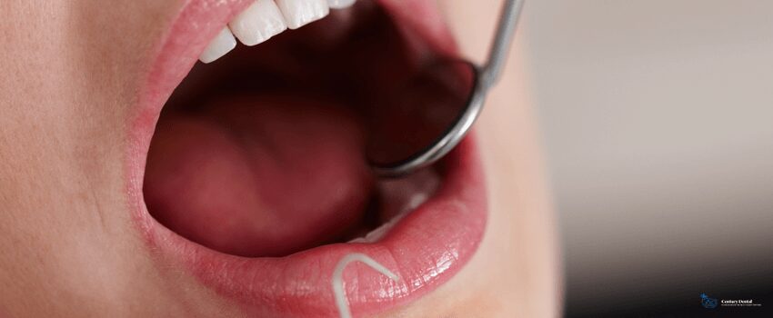
Staining Methods
Healthcare providers also use staining methods to identify erythroplakia. Fluorescence staining is a diagnostic method involving a special mouth rinse and a fluorescent light that shows abnormal areas.
Toluidine blue stain is another diagnostic method where erythroplakia lesions are coated with a blue dye. Areas that turn dark may indicate the presence of cancerous or precancerous cells.
Erythroplakia: Differential Diagnosis
The World Health Organization suggests health care providers consider other conditions before diagnosing erythroplakia. These conditions include:
- Erosive lichen planus
- Lupus erythematosus
- Hemangioma
- Pemphigus
- Acute atrophic candidiasis
- Nonhomogenous leukoplakia
How Is Erythroplakia Treated?
Since erythroplakia can progress into oral cancer, individuals with erythroplakia are advised to avoid known risk factors like alcohol and tobacco use. They should also practice good oral hygiene, visit their dentist regularly, and monitor their lesions to see if they resolve on their own.
Aside from frequent follow-ups, some treatment recommendations for erythroplakia include:
Laser Surgery
Laser therapies and surgeries involve focused light. Laser lights can be focused into powerful beams strong enough to cut steel or shape diamonds. In medicine, lasers allow surgeons to work with high precision on a small area, causing less pain and damage to surrounding tissues. However, it may result in scarring and swelling. It’s also expensive and may require multiple treatments.
Radiation Therapy
Radiation therapy uses high-energy X-rays and particles to kill cancerous cells or slow their growth. External beam radiotherapy is the most common type of radiation therapy used to treat oropharyngeal and oral cancers. It focuses radiation from an outside source onto the lesion or cancer. It feels more like taking an X-ray with a more potent dose. It’s painless and will only take a few minutes.
Radiation therapy in the mouth may damage your gums and teeth. See a dentist before you start treatment to ensure your oral health. They may recommend removing any bad teeth before you begin radiation treatment. They can also check and treat any oral issues that might arise during and after treatment.
Cryosurgery
Cryosurgery is a form of surgery that uses extremely cold temperatures to kill tumors and other abnormal tissues. This treatment often uses liquid nitrogen, which can instantly freeze almost anything it touches when it reaches a temperature between -346 to -320°F, instantly destroying cancerous cells. Cryosurgery can also treat skin tumors and lesions. It can also treat certain types of tumors inside the body.
Patients will need to follow future prevention strategies after a successful erythroplakia treatment. This includes avoiding risk factors, maintaining good oral hygiene, and life-long monitoring through regular visits to the dentist every 3-12 months. Some individuals may experience recurring lesions.
Key Takeaway
Erythroplakia is a rare condition that is characterized by red lesions that appear on your mucous membranes. Scraping these lesions causes them to bleed easily and can’t be classified as other conditions. They can transform into oral carcinoma if left untreated.
Dentists can diagnose erythroplakia as there are few symptoms other than irregular patches. They’ll likely recommend a biopsy to determine the presence of precancerous or cancerous cells. Heavy smoking, tobacco, and excessive alcohol consumption are some notable erythroplakia causes.
Treatment may involve a combination of lifestyle changes, such as quitting smoking and surgical removal.
Visit Century Dental today.
Early detection is important in treating precancerous lesions like erythroplakia. Dentists are often the first people to notice oral lesions like erythroplakia. Our dentist in St. Pete Beach can perform an oral cancer screening during a routine checkup. They can also perform other preventive dental services to help keep your teeth and gums healthy. Call us to schedule a check-up if you notice any erythroplakia symptoms and signs in your mouth.